Friday, 7 February 2020

PURETONE AUDIOMETRY


PURETONE AUDIOMETRY
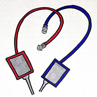
Audiometers are used to make quantitative measures of Air Conduction and Bone Conduction Pure Tone thresholds. AC thresholds assess the entire auditory pathway and are usually measured using earphones. When sound is delivered by an earphone, the hearing sensitivity can be assessed in each ear separately. BC thresholds are measured by placing a vibrator on the skull, with each ear assessed separately, usually by applying masking noise to the non test ear.  
Equipment
AUDIOMETERS
Puretones are generated within an audiometer. Audiometers have the ability to select tonal frequency and intensity level and to route tones to the left or right earphone. All audiometers also have an interrupter switch that presents the stimulus to the examinee.
The American National Standards Institute (ANSI) Specification for Audiometers (ANSI, 2010) describes four types of audiometers: Type 1 having the most features and Type 4 having the fewest features.
Type 1 audiometer
1.     Is a full-featured diagnostic audiometer.
2.     A Type 1 audiometer has earphones, bone vibrator, loud speakers, masking noise, and other features.
Type 4 audiometer
Is simply a screening device with earphones, but none of the other special features. Type 1 (full-featured, diagnostic audiometer) has the ability to assess puretone AC thresholds for frequencies ranging from 125 to 8,000 Hz and BC thresholds for frequencies ranging from 250 to 6,000 Hz. If an audiometer has extended high-frequency capability, air-conduction thresholds can be extended to 16,000 Hz. Maximum output levels for AC testing are as high as 120 dB HL for frequencies where hearing thresholds are most sensitive.  
Earphones
AYUSH SPEECH AND HEARING CLINIC

Earphones are generally used to test puretone AC thresholds. Supra-aural earphones, ones in which the cushion rests on the pinna, were the only choice for clinical audiology. The popularity of supra-aural phones was mainly due to their ease of calibration and the lack of other types of commercially available earphones. In the past few years, insert earphones and circumaural earphones have become available and provide some useful applications for puretone assessment.
Insert earphones are coupled to the ear by placing aprobe tip, typically a foam plug, into the ear canal. These earphones have gained popularity in the past few years because they offer distinct advantages over supra-aural earphones.
AYUSH SPEECH AND HEARING CLINIC

Advantages
Insert earphones yield higher levels of interaural attenuation than supra-aural earphones.
Interaural attenuation represents the decibel reduction of a sound as it crosses the head from the test ear to the nontest ear.
The average increase in interaural attenuation is roughly 20 dB. This reduces the need for masking the nontest ear and decreases the number of masking dilemmas, situations for which thresholds cannot be assessed, because the presentation level of the masking noise is possibly too high.
Another important advantage of insert earphones over supra-aural earphones is lower test–retest variability for thresholds obtained at 6 and 8 kHz; variability for other frequencies is comparable. Given that thresholds for 6 and 8 kHz are important for documenting changes in hearing due to noise exposure and for identifying acoustic tumors, lower variability should increase the diagnostic precision.
Insert earphones offer is elimination of collapsed ear canals. Supra-aural earphones cause the ear canal to narrow or be closed off entirely when the cushion presses against the pinna, collapsing the ear canal, resulting in false hearing thresholds, usually in the high frequencies. Because insert earphones keep the ear canal open, collapsed canals are eliminated.
Insert earphones is that they can be easily used with infants and toddlers who cannot or will not tolerate supra-aural earphones.
Insert earphones is the option of conducting middle-ear testing and otoacoustic emission testing without changing the earphones; some recently introduced diagnostic instruments use this approach. Although insert earphones offer a hygienic advantage over supra-aural earphones, because the foam tips that are placed into a client’s ear canal are disposable, the replacement cost of those tips is prohibitive for many applications. In addition to higher costs, insert earphones also yield errant thresholds in persons with eardrum perforations, including pressure-equalization tubes for additional
information about perforations.) Insert earphones also have maximum output levels that are lower than those produced by supra-aural earphones for some frequencies. Because of these differences, many diagnostic clinics keep both earphone types on hand and switch between them depending
on the application.
Speakers
AC thresholds can be measured using speakers as the transducer. Thresholds so obtained are known as sound-field thresholds. Sound-field thresholds are unable to provide ear-specific sensitivity estimates. In cases of unilateral hearing losses, the listener’s better ear determines threshold. This limitation and others dealing with control over stimulus level greatly limit clinical applications involving sound-field thresholds. Applications for sound-field thresholds are screening infant hearing or demonstrating to the parents their child’s hearing ability. Sound-field thresholds may also be desirable for a person wearing a hearing aid or cochlear implant. In sound-field threshold measures, the orientation of the listener to the speaker has a large effect on stimulus level presented at the eardrum. A person’s head and torso as well as the external ear affect sound levels. Differences in SPL at the eardrum are substantial for speaker locations at different distances and different angles relative to the listener. For this reason, sound-field calibration takes into consideration these factors. A mark is usually made on the ceiling (or floor) of the room to indicate the location of the listener during testing. Even at the desired location, stimulus level at the eardrum for some frequencies can vary as much as 20 dB or more by simply having the listener move his or her head. Calibration assumes the listener will always be facing the same direction relative to the sound source. Furniture and other persons in the sound field also affect the stimulus level at a listener’s eardrum. All of these factors add to the challenge of obtaining accurate sound-field thresholds. Another important consideration in sound-field
threshold measures is the stimulus type. Thresholds corresponding to different frequencies are desired for plotting an audiogram, but puretones can exhibit large differences in level at different positions in a testing suite as a result of standing waves. Standing waves occur when direct sound
from the speaker interacts with reflections, resulting in regions of cancellation and summation. Differences in stimulus level due to standing waves are minimized by using narrowband noise or frequency-modulated (FM) tones as the stimulus. FM tones, also known as warbled tones, are tones that vary in frequency over a range that is within a few percent of the nominal frequency. This variation occurs several times per second. Under earphones, thresholds obtained with these narrowband stimuli are nearly identical to thresholds obtained with puretones, with some exceptions in persons with steeply sloping hearing loss configurations. FM tones and narrowband noise are the preferred stimuli for sound-field threshold measures.
Bone Vibrators
AYUSH SPEECH AND HEARING CLINIC

A bone vibrator is a transducer that is designed to apply force to the skull when placed in contact with the head. Puretone BC thresholds are measured with a bone vibrator. A separation of 15 Db or more between masked AC and BC thresholds, with BC thresholds being lower than AC thresholds, is often evidence of a conductive hearing loss. Other possible explanations for air–bone gaps and bone–air gaps are equipment miscalibration, test–retest variability, and individual differences in anatomy that cause thresholds to deviate from the groupmean data used to derive normative values for relating AC
and BC thresholds. For threshold measurements bone vibrators are typically placed behind the pinna on the mastoid process or on the forehead. Placement on the mastoid process is preferred by 92% of audiologists. Mastoid placement is preferred mainly because it produces between 8 and 14 dB lower thresholds than forehead placement for the same power applied to the vibrator, depending on the frequency (ANSI, 2010). The median difference is 11.5 dB. Given that the maximum output limits for bone vibrators with mastoid placement are as much as 50 dB lower than that for AC thresholds, forehead placement yields an even larger difference. The inability to measure BC thresholds for higher levels means that a comparison of AC and BC thresholds is ambiguous in some cases. That is,
when BC thresholds indicate no response at the limits of the equipment and AC thresholds are poorer
than the levels where no response was obtained, the audiologist cannot establish from these thresholds whether the loss is purely sensory/neural or whether it has a conductive component.

Tuesday, 4 February 2020

Tuning Fork Tests


Tuning Fork Tests
AYUSH SPEECH AND HEARING CLINIC

Tuning forks were used to test hearing long before the development of the audiometer.
Schwabach Test
The Schwabach test is a technique for estimating a patient’s hearing sensitivity by bone-conduction. The test has two principal characteristics:
1.     It makes use of the fact that the tone produced by a tuning fork becomes softer with time after it has been struck due to damping, and
2.     The patient’s hearing is expressed in relative terms compared with the examiner’s hearing ability. This comparison is done by timing how long the tuning fork is heard by the patient and how long it is heard by the examiner. The basic procedure involves placing the base of the vibrating fork on the patient’s mastoid process until the tone fades away. The clinician then moves the fork to his own mastoid and times how long he can hear it. Compared with how long the examiner can hear the tone, it is expected that the patient will hear the tone
o    For a shorter period of time if she has a sensorineural loss;
o    For a longer period of time (or perhaps the same length of time) if she has a conductive loss; and
3.     For the same amount of time if she has normal hearing. Schwabach outcomes are problematic when dealing with mixed losses. The Schwabach test provides a relative estimate of hearing at best, and its validity is completely dependent on the tenuous assumption that the examiner really has normal hearing. It is not surprising this test is rarely if ever used.
Weber Test
The Weber test is used to help determine whether a unilateral hearing loss is sensorineural or conductive.
It is a lateralization test because the patient is asked to indicate the direction from which a sound
appears to be coming. Before starting this test, the patient should be advised that it is possible for the
tone to be heard from the good side or the poorer side, or any other location for that matter. The procedure involves putting the base of the vibrating tuning fork somewhere on the midline of the skull, most commonly on the center of the forehead or the top of the head. The audiometric Weber test uses the bone conduction vibrator instead of tuning forks. The patient is asked to indicate where the tone is heard. Hearing the tone in the better ear implies that there is a sensorineural loss in the poorer ear, whereas hearing the tone in the poorer ear suggests a conductive loss in that ear. The tone is heard in the middle of the head, “all over,” or equally in both ears when the patient has normal hearing, although some patients with sensorineural losses also report such midline lateralizations. If there is a mixed loss, the tone will be lateralized to the better ear if its level is below thepoorer ear’s bone-conduction threshold. The Weber test will fail to detect the conductive component of a mixed loss in such cases. The Weber test works for several reasons, all of which are related to the idea that the bone-conduction tone from the tuning fork reaches both cochleae at the same intensity.
The tone lateralizes to the better ear with sensorineural losses for either of two reasons:
1.     The tone will only be heard in the better ear if its level is lower than the bone-conduction threshold of the poorer ear.
2.     The second mechanism is due to the Stenger effect, which means that a sound presented to both ears is perceived only in the ear where it is louder. The intensity of the tone from the tuning fork will have a higher sensation level in the better ear than in the impaired ear. Hence, it will be louder in the better ear and will be perceived there. Several factors can explain why a bone-conduction tone would be louder in (and thus lateralized to) the poorer ear.  These mechanisms are just briefly mentioned because they are beyond the scope of an introductory text:
a.     Outer ear obstructions (e.g., impacted cerumen) may cause an occlusion effect,
b.    Mass loading of the middle ear system caused by effusions or ossicular chain interruptions may lower its resonance, and
c.     Phase advances may be caused by fixations or interruptions of the ossicular chain.
Bing Test
The Bing test is used to determine if closing off the patient’s ear canal results in an occlusion effect. The audiometric version of the test has already been discussed. In the traditional Bing test the patient is asked to report whether a tuning fork sounds louder with the ear canal open or closed. The base of a vibrating tuning fork is held against the patient’s mastoid process. The tester then presses the tragus down over the entrance of the ear canal to occlude it. The usual technique is to alternately occlude and unocclude the ear canal to help the patient make a reliable louder/softer judgment. It is desirable to make sure that the tuning fork sounds louder when the ear is closed and softer when the ear is open, instead of just asking whether the tone pulses between louder and softer. Unlike the audiometric version, which involves thresholds and thus quantifies the amount of the occlusion effect, the outcome of the tuning fork Bing test is based completely on a subjective judgment of louder versus not louder. If the occlusion effect is present, covering the ear canal should cause the tuning fork to sound louder. This is called a positive result and implies that the ear is either normal or has a sensorineural hearing loss. A negative result occurs if closing off the ear canal fails to make the tuning fork sound louder, and implies that there is either a conductive or mixed hearing loss.
Rinne Test
The Rinne test is a tuning fork procedure that compares hearing by air-conduction and by bone-conduction; however, the approach used is different from the one used in pure tone audiometry. The Rinne test is based on the idea that the hearing mechanism is normally more efficient by air-conduction than it is by bone-conduction. For this reason, a tuning fork will sound louder by air-conduction than by bone conduction. However, this air-conduction advantage is lost when there is a conductive hearing loss, in which case the tuning fork sounds louder by bone conduction than by air-conduction.
Administering the Rinne test involves asking the patient to indicate whether a vibrating tuning fork sounds louder when its base is held against the mastoid process (bone conduction) or when its prongs are held near the pinna, facing the opening of the ear canal (air-conduction). After striking the fork, the clinician alternates it between these two positions so that the patient can make a judgment about which one is louder. The bone-conduction vibrator is used instead of the tuning fork in the audiometric version of the Rinne test, and the patient indicates whether the vibrator sounds louder on the mastoid or in front of the ear canal. Masking noise must be put into the opposite ear to make sure that the Rinne results are really coming from the test ear.
The outcome of the Rinne test is traditionally called “positive” if the fork is louder by air-conduction, and this finding implies that the ear is normal or has a sensorineural hearing loss. The results are
called “negative” if bone-conduction is louder than air-conduction, which is interpreted as revealing the presence of a conductive abnormality. This terminology is confusing because the examiner is often concerned with identifying a conductive loss with this test. Consequently, many clinicians prefer to describe Rinne results as “air better than bone” (AC > BC) versus “bone better than air” (BC > AC). In these terms, AC > BC implies normal hearing or sensorineural impairment, and BC > AC implies a conductive disorder.
Sometimes, the air and bone-conduction signals sound equally loud to the patient (AC = BC). This equivocal outcome can usually be overcome by usingthe timed Rinne test (Gelfand 1977). This more accurate way to administer the Rinne test involves timing how long the patient can hear the tuning fork at the two locations.  
In this case, the results are Positive (AC > BC) when the tone is heard longer by air-conduction, and
a.     Negative (BC > AC) when it is heard longer by bone-conduction.
Another variation of the timed Rinne test involves holding the tuning fork at the mastoid until the tone has faded away, and then moving it to the ear canal (and vice versa).  Here, the result is
a.     AC > BC if the tone can still be heard by air-conduction after it faded away by boneconduction, and
b.    BC > AC if the tone can still be heard by bone-conduction after it faded away by air conduction.
Some Comments on Tuning Fork Tests
Tuning fork tests are quick and easy to administer and do not require special instrumentation, so they
can provide general, on-the-spot clinical insights, and they provide important (albeit limited) diagnostic information, especially when an audiogram is not available. However, tuning fork tests fall short of audiological measures in their ability to assess the patient’s hearing status. This is due to the greater
precision of audiometric tests made possible by calibrated electronic equipment and systematic testing strategies. Moreover, tuning fork tests are subject to considerable variability in administration and subjectivity in interpretation. Several limitations have already been mentioned with respect to individual tests, and others are worthy of mention. Which frequencies are tested varies among clinicians; some test only at 512 Hz and others use various combinations of frequencies. The intensity of the tone produced by the tuning fork depends on how hard it is struck each time, which causes stimulus levels to be inconsistent. Subjective patient responses can be a confounding variable, especially when dealing with younger children, and when the perception is “not logical” (e.g., when a tone is heard in the bad ear or gets louder when the ear is closed off). Also, tuning fork tests are usually done in examination rooms and clinics that are not sound isolated. Hence, noises that can mask the test signal and/or distract the patient are often a real issue.
It is therefore not surprising that carefully done
studies have shown that tuning forks are less accurate than audiometric methods. Wilson and Woods
(1975) found that both the Bing and Rinne tests failed to achieve a high level of accuracy in properly
identifying conductive versus nonconductive losses. Gelfand (1977) studied the diagnostic accuracy of the Rinne test. All of the tuning fork tests were done with masking of the opposite ear. He found that the Rinne test cannot identify a conductive loss with reasonable accuracy until the size of the air-bonegap becomes at least 25 to 40 dB wide. Thus, mild conductive hearing losses that are easily revealed audiometrically are frequently missed by tuning fork tests like the Rinne. Some tuning fork test problems are exacerbated by how and where the test is done. For example, the need for masking with certain tuning fork tests, particularly the Rinne. However, few physicians actually perform the Rinne test this way.

Friday, 31 January 2020

Noise-Induced Hearing Loss


Noise-Induced Hearing Loss


High sound levels can produce both temporary and permanent hearing losses due to over stimulation and/or mechanical trauma.  A sensorineural hearing loss produced by the damaging effects of over stimulation by high sound levels, usually over a long period of time, is called a noise induced hearing loss. In contrast, the term acoustic trauma usually refers to the hearing loss produced by extremely intense and impulsive sounds like explosions or gunshots. They can mechanically traumatize the eardrum, middle ear, and/or cochlear structures in addition to producing damage by over stimulation, and often from a single insult.
Almost everybody has experienced temporary hearing difficulty (often with tinnitus) after being exposed to high sound levels of one kind or another, such as loud music, construction noise, lawn mowers, subways, etc. This short-term decrease in hearing sensitivity is sensorineural in nature and is called a temporary threshold shift (TTS). In general, a TTS can be produced by sound levels greater than 80 dB sound pressure level (SPL). As the intensity and/or duration of the offending sound increases, the size of the TTS gets bigger and the time it takes for recovery gets longer. A permanent threshold shift (PTS) exists when the TTS does not recover completely, that is, when hearing sensitivity does not return to normal. Because PTS could refer to just about any permanent hearing loss, we generally lengthen the term to noise induced permanent threshold shift (NIPTS) for clarity. The nature and severity of a NIPTS is determined by the intensity, spectrum, duration, and time course of the offending sounds; the overall duration of the exposures over the years; and the patient’s individual susceptibility to the effects of noise. In addition, the amount of hearing loss produced by noise exposure is exacerbated if vibration is also present, and by the use of potentially ototoxic drugs.
The kinds of anatomical and physiological abnormalities caused by noise exposure range from the most subtle disruptions of hair cell metabolic activities and losses of stereocilia rigidity (leading to “floppy cilia”) to the complete degeneration of the organ of Corti and the auditory nerve supply. Both outer and inner hair cells are damaged by noise, but outer hair cells are more susceptible. Some of the abnormalities include metabolic exhaustion of the hair cells, structural changes and degeneration of structures within the hair cells, morphological changes of the cilia (so that they become fused and otherwise distorted), ruptures of cell membranes, and complete degeneration and loss of hair cells, neural cells, and supporting cells. Mild metabolic disruptions and floppy cilia can be reversible, and are thought to be related to TTS. It should be noted in this context that oxidative stress associated with accumulations of free radicals has been identified as a factor in noise-induced hearing loss Greater amounts of interference and damage are associated with permanent hearing losses.
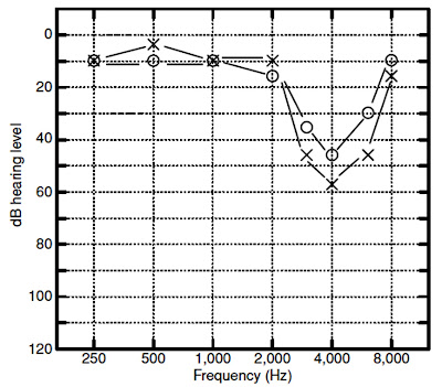
Unfortunately, noise exposures capable of producing temporary hearing loss can also cause permanent neural degeneration. Permanent degeneration of the auditory nerve cells even though the TTS was completely resolved and there was no loss of hair cells. Noise-induced impairments are usually associated with a notch-shaped high-frequency sensorineural loss that is worst at 4000 Hz although the notch often occurs at 3000 or 6000 Hz as well. The reason for the notch in this region is not definitively established. One explanation is that this region is most susceptible to damage due to the biology and mechanics of the cochlea. The cochlea with a boost in the 2000 to 4000 Hz region because of the resonance characteristics of the outer and middle ear. Noiseinduced losses tend to be bilateral and more or less symmetrical; however, there are many exceptions, especially when one ear has been subjected to more noise than the other. Not all “noise-induced” audiograms conform to the idealized picture in. Analyses of the progression of noise-induced hearing losses across many studies have revealed that the general audiometric pattern of noise-induced hearing loss evolves as noise exposure continues over the course of many years. The hearing loss typically begins as a notch at 4000 Hz. As noise exposure continues, the notch widens to include a wider range of frequencies, but continues to progress most noticeably at 4000 Hz. After perhaps 10 to 15 years of exposure, the progression of the loss at 4000 Hz often slows down, and progression now becomes more apparent at other frequencies, such as 2000 Hz.
WHY A NOTCH AT 4,000 HZ?
AYUSH SPEECH AND HEARING CLINIC

In humans, the frequency of maximum cochlear damage is one-half to one octave above the frequency of maximum stimulation. This phenomenon has to do with the angle of curvature of the human cochlea as well as less blood perfusion in the basal end of the cochlea compared to the apex. The human external ear (pinna and ear canal) influences the physical properties of sound outside the head (i.e., in the diffuse field) by resonating at frequencies between 2,000 and 4,000 Hz, depending on the volume and the length of the ear canal; for larger adult ears the maximum ear canal resonance, as measured with a probe microphone, is 2,600 to 3,000 Hz. In children, with shorter ear
canals with a smaller diameter, this ear canal resonance is higher in frequency. This resonance serves to amplify sound by 15 to 25 dB relative to the diffuse field (for instance, as measured at the shoulder) at the resonant frequency. Acousticians and engineers have referred to this resonance as the transfer function of the open ear (TFOE) or the external ear transfer function and is known to audiologists as the real ear–unaided response (REUR). When fitting hearing aids, placement of an earmold results in disruption of this normal ear canal resonance, resulting in insertion loss. The real ear–aided response (REAR) must provide amplification to compensate for the insertion loss, just to get back to the sound level that would arrive at the eardrum without the earmold or hearing aid in place. For broadband sound, the result of the TFOE (REUG) is an overall level measured at the eardrum roughly 7 dB higher than measured at the shoulder. Given that most environmental sound is relatively broadband, the frequency range of maximum stimulation is roughly one-half to one octave below 4,000 Hz. This is another reason why the 4,000-Hz frequency region is the most susceptible to damage.

Monday, 27 January 2020

EAR WAX


EAR WAX
AYUSH SPEECH AND HEARING CLINIC

An existing hearing loss can be exacerbated by the presence of ear wax. Ear wax can significantly reduce the transmission of sound by blocking the ear canal, blocking the sound
from exiting the hearing aid, or causing damage to internal components of the hearing aid.
What Is Ear Wax?
AYUSH SPEECH AND HEARING CLINIC

Ear wax is a normal product of the ear. Ear wax is primarily composed of keratin (dead skin) with a mixture of cerumen, sweat, dust, and other debris. The amount and consistency of ear wax vary from person to person. Ear wax can vary in color from yellow to orange or reddish-brown to dark brown or almost black. It may be nearly liquid or thick, sticky or dry, or soft or hard. Wax type is genetically inherited, although the appearance of wax may vary from time to time in the same person. Cerumen type has been used by anthropologists to track human migratory patterns, such as those of the Inuit. There are two main types, wet and dry. Dry flaky wax is common in persons of Asian descent and Native Americans. Dry wax contains by weight about 20% lipid. Wet wax is common in people of Western European descent (Caucasians) and people of African descent. Wet wax can be either soft or hard, with hard wax being more likely to be impacted.
Why Do We Have Ear Wax?
Various hypotheses have been advanced as to the purpose of ear wax. It has been proposed that wax provides protection against foreign objects, assists in cleaning the ear canal, acts as a lubricant, acts as an antibacterial and antifungal agent, and promotes a healthy immune response. Debris is removed from the ear canal by a “conveyor belt” process of epithelial migration that is aided by jaw movement. Cells of the tympanic membrane migrate outward from the umbo to the walls of the ear canal. The speed of cell migration accelerates as the cells move outward to the entrance of the ear canal. The cerumen in the canal is also carried outward, taking with it any dirt, dust, and particulate matter that may have gathered in the canal.
Wax can also act as a lubricant, preventing drying and itching of the skin in the ear canal (asteatosis). In wet-type cerumen, the lubricating effect is due to the presence of cholesterol, squalene, long-chain fatty acids, and alcohols produced by the sebaceous glands. Cerumen can provide protection against some strains of bacteria.
Removal of Ear Wax
If wax is hard and impacted in the ear canal, it may cause damage to the skin as it is removed and thus should be first softened. Wax removal is often more difficult for older people because their wax tends to be drier and harder. Ear wax can be softened by applying a few drops of mineral oil, baby oil, or glycerin in the ear for several days in a row. Oil should
be administered at night time so that it can be absorbed into the wax and skin overnight. If oil is administered in the morning, the oil will likely get into the hearing aid when inserted and possibly disable the hearing aid.
SYRINGING WITH WATER
AYUSH SPEECH AND HEARING CLINIC

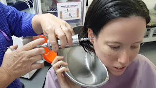
Syringing with water can be done by a client at home, by a trained audiologist, by a family doctor, or by another qualified person. Water pressure may, however, push the wax deeper into the canal (possibly touching the eardrum), whereas significant amounts of water may remain in the ear canal after syringing. When hydrogen peroxide (H2O2) is used, oxygen bubbles off, leaving water in the ear canal. A problem with wet, warm ear canals is that they make good incubators for growth of bacteria. In these instances, the ear canal may be flushed with isopropyl alcohol to displace the water and dry the skin but should be used sparingly to avoid excessive drying and itching.
PLASTIC SCOOPS
AYUSH SPEECH AND HEARING CLINIC

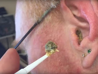
Small, flexible plastic scoops are commonly used by audiologists trained in wax removal. A good hands-free magnifier and light source are required. The basic technique is to gently scoop built-up wax from the canal. Care must be taken to minimize discomfort or trauma to the ear canal and to avoid contact with the tympanic membrane. This method is not recommended if wax is deeply impacted. Hairs in the ear canal may be embedded in the wax and can leave small amounts of blood in the canal when they are pulled out with the wax.
SUCTION
AYUSH SPEECH AND HEARING CLINIC

Suction is an effective way to remove wax and debris; however, there is a risk of damage to the ear canal and/or tympanic membrane. This method can be uncomfortable for the client, both physically because of the suction and acoustically because of the high SPLs. Suction should be used only by a qualified practitioner such as an otolaryngologist.
COTTON SWABS
AYUSH SPEECH AND HEARING CLINIC

Using cotton swabs to clean the ears is not recommended. Swabs tend to push wax deeper in the canal and may stimulate the production of more wax. Swabs irritate the skin of the ear canal and may damage the ear drum.
EAR CANDLING
AYUSH SPEECH AND HEARING CLINIC

Ear candling or coning is an ineffective and potentially dangerous method of cleaning the ears. A hollow candle is placed at the entrance of the ear canal and lit, supposedly sucking out ear wax. Despite many claims that ear candling is effective for wax removal, it has been proven that the substances appearing within the cone originate from the melted candle, not from the ears. The suction supposedly created by the candle’s flame is insufficient to remove wax and there is a substantial risk of burns, infection, obstruction of the ear canal, and perforation of the eardrum. Ear candling is not recommended at any time, and federal health warnings have been issued.
Cleaning Hearing Aids
AYUSH SPEECH AND HEARING CLINIC

Hearing aids should be cleaned regularly as a preventive measure. A thorough cleaning every 6 months is usually sufficient to reduce repairs due to wax damage. Some clients require deep cleaning of their hearing aids every month or even more frequently, whereas others may never have a problem with wax.
A vacuum chamber with a suction tip for cleaning hearing aids is essential for any hearing care practice. The vacuum chamber loosens and removes small particles of dust and wax, whereas the suction tip removes more recalcitrant debris. Care must be used when using a suction tip because the receiver can be easily damaged.
Prevention: The Use of Wax Guards Wax guards are the first line of defense against wax damage in a hearing aid. Different kinds of wax guards have been developed, including covers, metal springs, vented plastic plugs, and vented plastic baskets. One of the most effective is the vented plastic basket type, which is also the simplest for clients to change on their own. When clients cannot change the wax guard themselves, encourage them to bring their hearing aids in for regular cleaning and to change the wax guards.

INTELLECTUAL DISABILITY


INTELLECTUAL DISABILITY
AYUSH SPEECH AND HEARING CLINIC

The term intellectual disability includes impairments of general mental abilities that impact adaptive functioning. Symptoms of intellectual disability first appear during the developmental period and diagnosis requires a comprehensive assessment of intelligence across conceptual, social, and practical domains (American Psychiatric Association, 2013). Adaptive skill areas include:
·       Conceptual
·       Language
·       Reading
·       Writing
·       Math
·       Reasoning
·       Knowledge
·       Memory
·       Social
·       Empathy
·       Social judgment
·       Interpersonal communication skills
·       Ability to make and retain friendships
·       Practical/self-management
·       Personal care
·       Job responsibilities
·       Money management
·       Recreation
·       Organizing school and work tasks
Almost 10% of children with hearing loss also have intellectual disabilities. Those with an intellectual disability are at an increased risk for visual or hearing impairment or both. Detection and treatment of hearing loss in adults and children with intellectual disabilities is of utmost importance because hearing loss can exaggerate intellectual deficits by impeding the learning process. Down syndrome, also referred to as trisomy 21, is the leading cause of hearing loss and intellectual disabilities and occurs in approximately 1 in 700 births in the United States.  Audiologists are very likely to see a large number of children and adults with Down syndrome, a genetic disorder always associated with some degree of cognitive impairment. As individuals with Down syndrome age, there is a decline in intellectual ability. In fact, almost 100% of individuals with Down syndrome over 40 years of age demonstrate degenerative neuropathologic changes consistent with Alzheimer-type dementia.
Furthermore, some have speculated that the precocious aging of individuals with Down syndrome results in early presbycusis in this population. Hearing loss progresses more rapidly in adults with Down syndrome than those with other forms of intellectual disability or adults in the general population. Down syndrome is also frequently associated with conductive hearing loss and, less often, sensory/neural hearing loss. Although the majority of the conductive hearing losses in those with Down syndrome are secondary to middle ear effusion, some are the result of middle ear anomalies, such as ossicular malformations and damage to middle ear structures as a result of chronic infection. In contrast to the typically developing population, the prevalence of middle ear effusion tends to remain high in individuals with Down syndrome regardless of age. Found that adolescents with Down syndrome have poorer hearing and greater incidence of conductive hearing loss than their peers with intellectual disability, but without Down syndrome. For a comprehensive review of hearing loss associated with Down syndrome. 
Special Testing Considerations 
Little has been published on hearing assessment of adults with intellectual disability. However, it is well documented that audiologists must use test techniques that will bridge the difference between the chronologic and developmental age of individuals with cognitive disabilities to obtain valid test results. The patient’s mental or developmental age, not their chronologic age, should be considered when selecting appropriate test procedures and materials. Several investigators have evaluated the effectiveness of VRA with children having intellectual disabilities, including those with Down syndrome. With typically developing children and those with intellectual disabilities, VRA is effective with infants as young as 6 months cognitive developmental age. However, children with Down syndrome require a cognitive developmental age of 10 to 12 months to successfully participate in a VRA procedure. Furthermore, behavioral thresholds of infants with Down syndrome have been found to be 10 to 25 dB poorer than those of typically developing infants when all had normal hearing verified via ABR. This elevation of behavioural thresholds is presumed to be the result of more inattentive behavior on the part of the children with Down syndrome relative to their typically developing peers. Moreover, this inattentive behavior provides additional reason to utilize a test battery that includes physiological measures when testing children with Down syndrome. Although it is recommended that audiologists attempt to elicit a spontaneous head-turn response during the VRA conditioning process, some children with intellectual disability may not have developed auditory localization ability. Recall that auditory localization is a higher order skill than detection, the required skill for VRA. In such cases, several administrations of paired conditioning trials may be required. If the patient does not respond to the auditory stimuli, the audiologist may be left with the question, “Does the patient not hear the stimuli, or can she or he not perform the task?” One method that can answer this question is for the audiologist to place the bone vibrator either in the patient’s hand or on the head and, using a low-frequency stimulus at approximately 50 to 60 dB hearing level (HL), determine if the patient can perform the task using this vibrotactile cue. In this way, the patient is able to feel the stimulus and, thus, is not required to hear to participate. If the patient is able to cooperate for the task under these vibrotactile conditions, then the audiologist should return to the auditory stimuli and continue testing with the knowledge that the patient understands the task. If using a play audiometric technique, it is often appropriate for the audiologist to demonstrate the play task to the patient with intellectual disability rather than attempting to explain the instructions verbally. Because learning the desired response behaviors may take longer for children and adults with intellectual disability, it may be useful to have them practice the listening task at home before coming to the clinic.