Friday 31 January 2020

Noise-Induced Hearing Loss


Noise-Induced Hearing Loss


High sound levels can produce both temporary and permanent hearing losses due to over stimulation and/or mechanical trauma.  A sensorineural hearing loss produced by the damaging effects of over stimulation by high sound levels, usually over a long period of time, is called a noise induced hearing loss. In contrast, the term acoustic trauma usually refers to the hearing loss produced by extremely intense and impulsive sounds like explosions or gunshots. They can mechanically traumatize the eardrum, middle ear, and/or cochlear structures in addition to producing damage by over stimulation, and often from a single insult.
Almost everybody has experienced temporary hearing difficulty (often with tinnitus) after being exposed to high sound levels of one kind or another, such as loud music, construction noise, lawn mowers, subways, etc. This short-term decrease in hearing sensitivity is sensorineural in nature and is called a temporary threshold shift (TTS). In general, a TTS can be produced by sound levels greater than 80 dB sound pressure level (SPL). As the intensity and/or duration of the offending sound increases, the size of the TTS gets bigger and the time it takes for recovery gets longer. A permanent threshold shift (PTS) exists when the TTS does not recover completely, that is, when hearing sensitivity does not return to normal. Because PTS could refer to just about any permanent hearing loss, we generally lengthen the term to noise induced permanent threshold shift (NIPTS) for clarity. The nature and severity of a NIPTS is determined by the intensity, spectrum, duration, and time course of the offending sounds; the overall duration of the exposures over the years; and the patient’s individual susceptibility to the effects of noise. In addition, the amount of hearing loss produced by noise exposure is exacerbated if vibration is also present, and by the use of potentially ototoxic drugs.
The kinds of anatomical and physiological abnormalities caused by noise exposure range from the most subtle disruptions of hair cell metabolic activities and losses of stereocilia rigidity (leading to “floppy cilia”) to the complete degeneration of the organ of Corti and the auditory nerve supply. Both outer and inner hair cells are damaged by noise, but outer hair cells are more susceptible. Some of the abnormalities include metabolic exhaustion of the hair cells, structural changes and degeneration of structures within the hair cells, morphological changes of the cilia (so that they become fused and otherwise distorted), ruptures of cell membranes, and complete degeneration and loss of hair cells, neural cells, and supporting cells. Mild metabolic disruptions and floppy cilia can be reversible, and are thought to be related to TTS. It should be noted in this context that oxidative stress associated with accumulations of free radicals has been identified as a factor in noise-induced hearing loss Greater amounts of interference and damage are associated with permanent hearing losses.
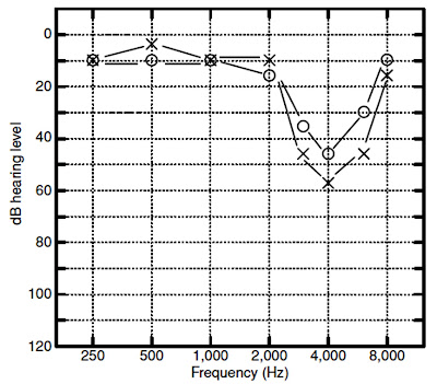
Unfortunately, noise exposures capable of producing temporary hearing loss can also cause permanent neural degeneration. Permanent degeneration of the auditory nerve cells even though the TTS was completely resolved and there was no loss of hair cells. Noise-induced impairments are usually associated with a notch-shaped high-frequency sensorineural loss that is worst at 4000 Hz although the notch often occurs at 3000 or 6000 Hz as well. The reason for the notch in this region is not definitively established. One explanation is that this region is most susceptible to damage due to the biology and mechanics of the cochlea. The cochlea with a boost in the 2000 to 4000 Hz region because of the resonance characteristics of the outer and middle ear. Noiseinduced losses tend to be bilateral and more or less symmetrical; however, there are many exceptions, especially when one ear has been subjected to more noise than the other. Not all “noise-induced” audiograms conform to the idealized picture in. Analyses of the progression of noise-induced hearing losses across many studies have revealed that the general audiometric pattern of noise-induced hearing loss evolves as noise exposure continues over the course of many years. The hearing loss typically begins as a notch at 4000 Hz. As noise exposure continues, the notch widens to include a wider range of frequencies, but continues to progress most noticeably at 4000 Hz. After perhaps 10 to 15 years of exposure, the progression of the loss at 4000 Hz often slows down, and progression now becomes more apparent at other frequencies, such as 2000 Hz.
WHY A NOTCH AT 4,000 HZ?
AYUSH SPEECH AND HEARING CLINIC

In humans, the frequency of maximum cochlear damage is one-half to one octave above the frequency of maximum stimulation. This phenomenon has to do with the angle of curvature of the human cochlea as well as less blood perfusion in the basal end of the cochlea compared to the apex. The human external ear (pinna and ear canal) influences the physical properties of sound outside the head (i.e., in the diffuse field) by resonating at frequencies between 2,000 and 4,000 Hz, depending on the volume and the length of the ear canal; for larger adult ears the maximum ear canal resonance, as measured with a probe microphone, is 2,600 to 3,000 Hz. In children, with shorter ear
canals with a smaller diameter, this ear canal resonance is higher in frequency. This resonance serves to amplify sound by 15 to 25 dB relative to the diffuse field (for instance, as measured at the shoulder) at the resonant frequency. Acousticians and engineers have referred to this resonance as the transfer function of the open ear (TFOE) or the external ear transfer function and is known to audiologists as the real ear–unaided response (REUR). When fitting hearing aids, placement of an earmold results in disruption of this normal ear canal resonance, resulting in insertion loss. The real ear–aided response (REAR) must provide amplification to compensate for the insertion loss, just to get back to the sound level that would arrive at the eardrum without the earmold or hearing aid in place. For broadband sound, the result of the TFOE (REUG) is an overall level measured at the eardrum roughly 7 dB higher than measured at the shoulder. Given that most environmental sound is relatively broadband, the frequency range of maximum stimulation is roughly one-half to one octave below 4,000 Hz. This is another reason why the 4,000-Hz frequency region is the most susceptible to damage.

Monday 27 January 2020

EAR WAX


EAR WAX
AYUSH SPEECH AND HEARING CLINIC

An existing hearing loss can be exacerbated by the presence of ear wax. Ear wax can significantly reduce the transmission of sound by blocking the ear canal, blocking the sound
from exiting the hearing aid, or causing damage to internal components of the hearing aid.
What Is Ear Wax?
AYUSH SPEECH AND HEARING CLINIC

Ear wax is a normal product of the ear. Ear wax is primarily composed of keratin (dead skin) with a mixture of cerumen, sweat, dust, and other debris. The amount and consistency of ear wax vary from person to person. Ear wax can vary in color from yellow to orange or reddish-brown to dark brown or almost black. It may be nearly liquid or thick, sticky or dry, or soft or hard. Wax type is genetically inherited, although the appearance of wax may vary from time to time in the same person. Cerumen type has been used by anthropologists to track human migratory patterns, such as those of the Inuit. There are two main types, wet and dry. Dry flaky wax is common in persons of Asian descent and Native Americans. Dry wax contains by weight about 20% lipid. Wet wax is common in people of Western European descent (Caucasians) and people of African descent. Wet wax can be either soft or hard, with hard wax being more likely to be impacted.
Why Do We Have Ear Wax?
Various hypotheses have been advanced as to the purpose of ear wax. It has been proposed that wax provides protection against foreign objects, assists in cleaning the ear canal, acts as a lubricant, acts as an antibacterial and antifungal agent, and promotes a healthy immune response. Debris is removed from the ear canal by a “conveyor belt” process of epithelial migration that is aided by jaw movement. Cells of the tympanic membrane migrate outward from the umbo to the walls of the ear canal. The speed of cell migration accelerates as the cells move outward to the entrance of the ear canal. The cerumen in the canal is also carried outward, taking with it any dirt, dust, and particulate matter that may have gathered in the canal.
Wax can also act as a lubricant, preventing drying and itching of the skin in the ear canal (asteatosis). In wet-type cerumen, the lubricating effect is due to the presence of cholesterol, squalene, long-chain fatty acids, and alcohols produced by the sebaceous glands. Cerumen can provide protection against some strains of bacteria.
Removal of Ear Wax
If wax is hard and impacted in the ear canal, it may cause damage to the skin as it is removed and thus should be first softened. Wax removal is often more difficult for older people because their wax tends to be drier and harder. Ear wax can be softened by applying a few drops of mineral oil, baby oil, or glycerin in the ear for several days in a row. Oil should
be administered at night time so that it can be absorbed into the wax and skin overnight. If oil is administered in the morning, the oil will likely get into the hearing aid when inserted and possibly disable the hearing aid.
SYRINGING WITH WATER
AYUSH SPEECH AND HEARING CLINIC

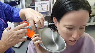
Syringing with water can be done by a client at home, by a trained audiologist, by a family doctor, or by another qualified person. Water pressure may, however, push the wax deeper into the canal (possibly touching the eardrum), whereas significant amounts of water may remain in the ear canal after syringing. When hydrogen peroxide (H2O2) is used, oxygen bubbles off, leaving water in the ear canal. A problem with wet, warm ear canals is that they make good incubators for growth of bacteria. In these instances, the ear canal may be flushed with isopropyl alcohol to displace the water and dry the skin but should be used sparingly to avoid excessive drying and itching.
PLASTIC SCOOPS
AYUSH SPEECH AND HEARING CLINIC

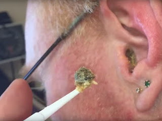
Small, flexible plastic scoops are commonly used by audiologists trained in wax removal. A good hands-free magnifier and light source are required. The basic technique is to gently scoop built-up wax from the canal. Care must be taken to minimize discomfort or trauma to the ear canal and to avoid contact with the tympanic membrane. This method is not recommended if wax is deeply impacted. Hairs in the ear canal may be embedded in the wax and can leave small amounts of blood in the canal when they are pulled out with the wax.
SUCTION
AYUSH SPEECH AND HEARING CLINIC

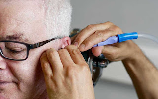
Suction is an effective way to remove wax and debris; however, there is a risk of damage to the ear canal and/or tympanic membrane. This method can be uncomfortable for the client, both physically because of the suction and acoustically because of the high SPLs. Suction should be used only by a qualified practitioner such as an otolaryngologist.
COTTON SWABS
AYUSH SPEECH AND HEARING CLINIC

Using cotton swabs to clean the ears is not recommended. Swabs tend to push wax deeper in the canal and may stimulate the production of more wax. Swabs irritate the skin of the ear canal and may damage the ear drum.
EAR CANDLING
AYUSH SPEECH AND HEARING CLINIC

Ear candling or coning is an ineffective and potentially dangerous method of cleaning the ears. A hollow candle is placed at the entrance of the ear canal and lit, supposedly sucking out ear wax. Despite many claims that ear candling is effective for wax removal, it has been proven that the substances appearing within the cone originate from the melted candle, not from the ears. The suction supposedly created by the candle’s flame is insufficient to remove wax and there is a substantial risk of burns, infection, obstruction of the ear canal, and perforation of the eardrum. Ear candling is not recommended at any time, and federal health warnings have been issued.
Cleaning Hearing Aids
AYUSH SPEECH AND HEARING CLINIC

Hearing aids should be cleaned regularly as a preventive measure. A thorough cleaning every 6 months is usually sufficient to reduce repairs due to wax damage. Some clients require deep cleaning of their hearing aids every month or even more frequently, whereas others may never have a problem with wax.
A vacuum chamber with a suction tip for cleaning hearing aids is essential for any hearing care practice. The vacuum chamber loosens and removes small particles of dust and wax, whereas the suction tip removes more recalcitrant debris. Care must be used when using a suction tip because the receiver can be easily damaged.
Prevention: The Use of Wax Guards Wax guards are the first line of defense against wax damage in a hearing aid. Different kinds of wax guards have been developed, including covers, metal springs, vented plastic plugs, and vented plastic baskets. One of the most effective is the vented plastic basket type, which is also the simplest for clients to change on their own. When clients cannot change the wax guard themselves, encourage them to bring their hearing aids in for regular cleaning and to change the wax guards.

INTELLECTUAL DISABILITY


INTELLECTUAL DISABILITY
AYUSH SPEECH AND HEARING CLINIC

The term intellectual disability includes impairments of general mental abilities that impact adaptive functioning. Symptoms of intellectual disability first appear during the developmental period and diagnosis requires a comprehensive assessment of intelligence across conceptual, social, and practical domains (American Psychiatric Association, 2013). Adaptive skill areas include:
·       Conceptual
·       Language
·       Reading
·       Writing
·       Math
·       Reasoning
·       Knowledge
·       Memory
·       Social
·       Empathy
·       Social judgment
·       Interpersonal communication skills
·       Ability to make and retain friendships
·       Practical/self-management
·       Personal care
·       Job responsibilities
·       Money management
·       Recreation
·       Organizing school and work tasks
Almost 10% of children with hearing loss also have intellectual disabilities. Those with an intellectual disability are at an increased risk for visual or hearing impairment or both. Detection and treatment of hearing loss in adults and children with intellectual disabilities is of utmost importance because hearing loss can exaggerate intellectual deficits by impeding the learning process. Down syndrome, also referred to as trisomy 21, is the leading cause of hearing loss and intellectual disabilities and occurs in approximately 1 in 700 births in the United States.  Audiologists are very likely to see a large number of children and adults with Down syndrome, a genetic disorder always associated with some degree of cognitive impairment. As individuals with Down syndrome age, there is a decline in intellectual ability. In fact, almost 100% of individuals with Down syndrome over 40 years of age demonstrate degenerative neuropathologic changes consistent with Alzheimer-type dementia.
Furthermore, some have speculated that the precocious aging of individuals with Down syndrome results in early presbycusis in this population. Hearing loss progresses more rapidly in adults with Down syndrome than those with other forms of intellectual disability or adults in the general population. Down syndrome is also frequently associated with conductive hearing loss and, less often, sensory/neural hearing loss. Although the majority of the conductive hearing losses in those with Down syndrome are secondary to middle ear effusion, some are the result of middle ear anomalies, such as ossicular malformations and damage to middle ear structures as a result of chronic infection. In contrast to the typically developing population, the prevalence of middle ear effusion tends to remain high in individuals with Down syndrome regardless of age. Found that adolescents with Down syndrome have poorer hearing and greater incidence of conductive hearing loss than their peers with intellectual disability, but without Down syndrome. For a comprehensive review of hearing loss associated with Down syndrome. 
Special Testing Considerations 
Little has been published on hearing assessment of adults with intellectual disability. However, it is well documented that audiologists must use test techniques that will bridge the difference between the chronologic and developmental age of individuals with cognitive disabilities to obtain valid test results. The patient’s mental or developmental age, not their chronologic age, should be considered when selecting appropriate test procedures and materials. Several investigators have evaluated the effectiveness of VRA with children having intellectual disabilities, including those with Down syndrome. With typically developing children and those with intellectual disabilities, VRA is effective with infants as young as 6 months cognitive developmental age. However, children with Down syndrome require a cognitive developmental age of 10 to 12 months to successfully participate in a VRA procedure. Furthermore, behavioral thresholds of infants with Down syndrome have been found to be 10 to 25 dB poorer than those of typically developing infants when all had normal hearing verified via ABR. This elevation of behavioural thresholds is presumed to be the result of more inattentive behavior on the part of the children with Down syndrome relative to their typically developing peers. Moreover, this inattentive behavior provides additional reason to utilize a test battery that includes physiological measures when testing children with Down syndrome. Although it is recommended that audiologists attempt to elicit a spontaneous head-turn response during the VRA conditioning process, some children with intellectual disability may not have developed auditory localization ability. Recall that auditory localization is a higher order skill than detection, the required skill for VRA. In such cases, several administrations of paired conditioning trials may be required. If the patient does not respond to the auditory stimuli, the audiologist may be left with the question, “Does the patient not hear the stimuli, or can she or he not perform the task?” One method that can answer this question is for the audiologist to place the bone vibrator either in the patient’s hand or on the head and, using a low-frequency stimulus at approximately 50 to 60 dB hearing level (HL), determine if the patient can perform the task using this vibrotactile cue. In this way, the patient is able to feel the stimulus and, thus, is not required to hear to participate. If the patient is able to cooperate for the task under these vibrotactile conditions, then the audiologist should return to the auditory stimuli and continue testing with the knowledge that the patient understands the task. If using a play audiometric technique, it is often appropriate for the audiologist to demonstrate the play task to the patient with intellectual disability rather than attempting to explain the instructions verbally. Because learning the desired response behaviors may take longer for children and adults with intellectual disability, it may be useful to have them practice the listening task at home before coming to the clinic.

Thursday 23 January 2020

PHYSICAL DISABILITIES


PHYSICAL DISABILITIES
AYUSH SPEECH AND HEARING CLINIC
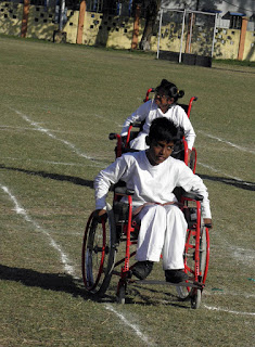
Persons who are deaf or hard of hearing should have similar motor development and skills as those with normal hearing unless vestibular function is affected. That is, deafness alone does not affect motor abilities or balance function. In fact, 93% of children with deafness have average to above average motor skills. Environmental factors such as emphasis on physical skills in the school curriculum, opportunities for practice and play, and parenting styles are believed to influence physical development of children with hearing loss. Audiologists should be aware of expected gross motor milestones in typically developing children. If a child with hearing loss is not walking by 15 months of age, a referral for further evaluation by a developmental psychologist or pediatrician is warranted.
Vestibular abnormalities that can result in gross motor problems include cochlear malformations such as Mondini’s deformity and cochlear hypoplasia. Other congenital causes of gross motor deficits in children with hearing loss include syndromes such as CHARGE syndrome and Usher syndrome type I (described in a later section) and CP. CP is a disorder of neuromotor function. Approximately 3% of children with hearing loss also have been diagnosed with CP, which is characterized by an inability to control motor function as a result of damage to or an anomaly of the developing brain. This damage interferes with messages from the brain to the body and from the body to the brain. The effects of CP vary widely from individual to individual. There are three primary types of CP:
·    Spastic—characterized by high muscle tone (hypertonia) producing stiff and difficult movement
·    Athetoid—producing involuntary and uncontrolled movement
·   Ataxic—characterized by low muscle tone (hypotonia) producing a disturbed sense of balance, disturbed position in space, and general uncoordinated movement
·    Quadriplegia—all four limbs are involved
·    Diplegia—all four limbs are involved and both legs are more severely affected than the arms
·    Hemiplegia—one side of the body is affected and the arm is usually more involved than the leg
·    Triplegia—three limbs are involved, usually both arms and a leg
·    Monoplegia—only one limb is affected, usually an arm
CP is not a progressive condition. The damage to the brain is a one-time event. However, the effects may change over time. For example, with physical therapy a child’s gross and fine motor skills may improve with time. However, the aging process can be harder on bodies with abnormal posture or that have had little exercise, so the effects may result in a gradual decline in motoric ability. It is important to remember that the degree of physical disability experienced by a person with CP is not an indication of his or her level of intelligence.
The brain damage that caused CP may also lead to other conditions such as learning disabilities or developmental delays. Approximately 20% of children with CP will also experience hearing or language problems. The hearing loss is typically sensory/neural in nature. In addition, between 40% and 75% of individuals with CP will also have some degree of vision deficit.
Special Testing Considerations
Individuals with motor delays may not respond behaviourally to auditory stimuli because their physical disabilities limit their ability to orient to sound. However, when testing children, VRA can still provide reliable information even for those with poor head and neck control. Modifications that might need to be made in the test arrangements for VRA include the use of an infant seat to provide additional head support. However, audiologists should ensure that head supports do not block the ears and impede sound field stimuli. If children with motor difficulties cannot make a head-turn response to sound, response modifications can be made. Modifications include alternative responses such as localizing to the sound stimuli with their eyes as opposed to head turns. CPA might also require modifications. Response modifications might need to include options that do not require the use of fine motor skills. Examples of such modifications could include asking a child to drop a ball into a large bucket rather than having the child insert a peg in a pegboard, partial hand raising, or even just a head nod. Additionally, a variety of gross motor responses can be used to trigger an electronic switch that will, in turn, activate a computer screen programmed for appropriate visual reinforcement.
If the physical disability has a neuromotor component, such as with CP, physiological measures might be affected. That is, abnormality in measures such as the auditory brainstem response (ABR) may be misinterpreted as indicative of hearing loss when, in fact, the abnormality is in neurotransmission. Therefore, interpretation of the ABR must be made cautiously and in concert
with the entire battery of auditory tests, behavioral and physiological. Sedation may be required when conducting ABR with individuals who have CP in an attempt to relax their head and neck or to reduce extraneous muscle movements, thus reducing myogenic artifact.

Wednesday 22 January 2020

AUTISM SPECTRUM DISORDER


AUTISM SPECTRUM DISORDER

AYUSH SPEECH AND HEARING CLINIC
Autism spectrum disorder (ASD) is a developmental disorder characterized by symptoms appearing in early childhood and impairing day-to-day life function. These symptoms include qualitative impairments in social/communication interaction and repetitive and restricted behaviors, according to the Diagnostic and Statistics Manual of Mental Disorders. Under the umbrella of ASD, a patient’s symptoms will fall on a continuum, with some showing mild symptoms and others, more severe. A diagnosis under the general diagnostic category of ASD is relatively new. Prior to the publication of DSM-5, there were five ASDs, each of which had a unique diagnosis: classic autism, pervasive developmental disorder (PDD), Asperger’s disorder, Rett’s syndrome, and childhood disintegrative disorder. With the exception of Rett’s syndrome, these disorders are now subsumed into the diagnosis of ASD. Rett’s syndrome is now its own entity and is no longer a part of the autism spectrum.
ASD is thought to have an early onset, with symptoms appearing before 24 months of age in most cases. Although a definitive diagnosis of autism is not generally made until the age of 3 years or later (Mandell et al., 2005), there are a growing number of reports of stable diagnoses following identification as young as 2 years (Chawarska et al., 2009). Prevalence estimates of ASD have increased steadily over time from reports of 1 to 5 children per 10,000 in the 1970s. Current numbers from the Centers for Disease Control and Prevention suggest a prevalence of 114 per 10,000 children (Baio, 2012; Rice, 2009). It remains to be seen whether there has been a true increase in prevalence of ASD over time or the reported changes in prevalence can be explained by changes in diagnostic criteria and increased awareness of the disorder by parents and professionals. Boys are more likely to be affected with autism than girls, at a ratio of more than 3:1. About 50% to 70% of children with ASD also have an intellectual disability.
There is no strong evidence to suggest that individuals with ASD have a greater risk of hearing loss than the general population. However, the presence of unusual sensory responses, including abnormal responses to sound, is considered an associated feature of ASD. For example, individuals with ASD might completely ignore sounds that would result in a reaction from typically developing individuals. Other times, they often appear to be overly sensitive to sound by covering their ears with their hands when loud or unexpected sounds occur. In addition to these abnormal responses to sound, young children with ASD are known to lag behind on language milestones. Therefore, those with ASD will likely be referred to audiologists for hearing assessments as part of the developmental evaluation to rule out hearing loss as the cause of language delay. On average, behavioral responses to sound of children with ASD who have normal hearing are elevated and less reliable relative to those of typically developing children. Relatively little is known about higher order auditory abilities of individuals with ASD. However, altered temporal processing has been recorded in both adults and children with ASD.
AYUSH SPEECH AND HEARING CLINIC

Special Testing Considerations
Children with ASD who have hearing loss are diagnosed, on average, almost 1 year later than those without hearing loss. Therefore, it is reasonable for audiologists to be alert to the general behavioral characteristics of childhood ASD to facilitate referral for evaluation when indicated. Several screening tools are available that can be used by audiologists. These include, among others, the Modified Checklist for Autism in Toddlers (M-CHAT) and the Pervasive Developmental Disorder Screening Test II (PDDST-II).
Understanding the general behavioral characteristics of those with ASD can also be helpful to audiologists as they consider modifications to the traditional test battery. Because the majority of those with ASD exhibit cognitive deficits, behavioural abnormalities, and hypersensitivity to sensory stimulation, audiologists should be prepared to address those issues during the test session. For instance, transitions are often difficult for individuals with ASD. When possible, audiologists should avoid travel from room to room with the patient, taking care to escort the patient to the testing area immediately rather than keeping him or her in the waiting area.
Regardless of the chronologic age of the individuals, audiologists will need to use behavioral test procedures that are appropriate for their patient’s cognitive level. This may mean that procedures typically used with infants and young children such as visual reinforcement audiometry (VRA) or play audiometric techniques will be used with older children or even adults. If VRA is used, one should consider minimizing the impact of the reinforcement by turning off the animation (if a lighted, animated toy is used) or using a video reinforcement. Other testing options for patients functioning at a developmental level of 2.5 years or greater are conditioned play audiometry (CPA) and tangible-reinforcement operant conditioning audiometry (TROCA). TROCA is often used in pediatric practices that specialize in serving those with multiple disabilities. TROCA requires the patient to press a bar or a button whenever a sound is heard, which is paired with the dispensing of a tangible reinforcement (e.g., small piece of food). TROCA is noted to be particularly effective with children having cognitive or behavioral (e.g., ASD) disorders. A significant number of children with ASD receive other clinical services (e.g., speech therapy).
Patients with ASD are often resistant to earphones or probes used for individual ear testing. Audiologists can ask the parent or caregiver to practice listening activities with headphones with the patient prior to the appointment. If a patient with ASD will not allow the placement of earphones or probes, audiologists might have to resort to sedated procedures. This is certainly true if one plans to fit hearing aids. Individuals with ASD are known to be difficult to sedate with currently available pediatric sedating agents and are at risk for seizures while under sedation. Therefore, consultation with the physician in charge of administering and monitoring the sedation process will need to include notification of the patient’s diagnosis of ASD.

Tuesday 21 January 2020

HYPERACUSIS


HYPERACUSIS

AYUSH SPEECH AND HEARING CLINIC

Introduction
Hyperacusis can involve loudness, annoyance, fear, and pain. We have noted that tinnitus is often accompanied by hyperacusis, and many current sound therapy protocols treat tinnitus and hyperacusis in parallel. One can readily imagine that sounds perceived as being very loud could easily become annoying. The anticipation of loud and/or annoying sounds could reasonably lead to the fear of these sounds. However, it is possible for sounds to be annoying or feared without being too loud. Patients also report that some sounds are physically painful, usually those perceived as loud. Occasionally, patients with tinnitus report that some sounds make their tinnitus worse. It is important to separate each of these symptoms, both for the patient and the clinician, to understand the problems carefully, and to offer treatment suggestions.
Neurophysiological Causes, Mechanisms, and Models of Hyperacusis
Anything that which causes a sensory/neural hearing loss can likely also cause hyperacusis. Hyperacusis can also occur without identifiable hearing loss. As a stimulus is increased, the activity of individual nerve fibers increases, and the number of nerve fibers activated increases (and usually its perceived loudness also increases). Moderately intense sounds might result in loudness hyperacusis if
1. greater than normal activity was produced on individual nerve fibers,
2. more nerve fibers were activated than normal, and/or
3. there was greater than normal synchrony across fibers
We suggest that hyperacusis might also be a function of such brain plasticity. Following a peripheral hearing loss, say at 4,000 Hz, nerve fibers in the brain that normally respond to 4,000 Hz begin to respond to other, nearby frequencies, for example, 3,000 Hz. This results in more nerve fibers in the brain responding to 3,000 Hz than would be present normally. If hyperacusis is related to the number of fibers activated, this could account for it as a phenomenon. Hazell (1987) suggested that hyperacusis might be the result of an “abnormal gain control.” It is as if the brain receives a lack of information after hearing loss and therefore turns up some hypothetical gain control.
EVALUATION OF HYPERACUSIS
Medical
The medical evaluation for hyperacusis parallels that for tinnitus. Some conditions have been associated with hyperacusis, including facial paralysis, head trauma, and metabolic disorders, infections (Lyme disease), and genetic (Williams’ syndrome) abnormalities.
Measuring Hyperacusis
LOUDNESS HYPERACUSIS
Loudness Discomfort Levels
Loudness discomfort levels (LDLs) can be performed with puretones at 500 and 4,000 Hz in each ear. We use the following instructions: “This is a test in which you will be hearing sounds in your right/left ear. We want you to decide when the sound first becomes uncomfortably loud.”
Magnitude Estimation of Loudness
It is possible to present tones and ask for a rating of loudness on a scale from 0 to 100, with 100 being the loudest sound a person can imagine. Hyperacusis scales have been developed to attempt
to differentiate loudness and annoyance and to ascertain a general idea of the impact of hyperacusis on a patient’s daily activities. The questionnaire asks individuals to consider several typical events they might encounter in their daily lives. They then separately rate the loudness and the annoyance for the same situations. For example, a patient may rate “telephone ringing in the same room” as 40 out of 100 on the loudness scale (with 100 being unbearably loud), whereas rating it as 85 out of 100 on the annoyance scale (with 100 being unbearably annoying).
ANNOYANCE HYPERACUSIS
In terms of hearing loss tinnitus, and hyperacusis the statement include items such as ‘you avoid shopping’ ‘you feel depressed’ and allow clinician to separate the impact on function that patient perceive from where hearing loss, tinnitus and hyperacusis. Another approach we have tried is to have patients rate recorded sounds. For example, we have patients rate recorded sounds of dishes hitting together, a lawn mower, and crowd noise. A multiple activity scale for annoyance hyperacusis, providing 15 situations (e.g., concert, shopping center, work, church, children). Subjects rated from 1 to 10 each of the “relevant” activities, which were averaged for a total score. They also had patients rate annoyance hyperacusis on a scale from1 to 10.
FEAR HYPERACUSIS
Patients can develop a fear of very specific sounds or categories of sounds (e.g., those containing high frequencies) or of any intense sound. The simplest approach may be to ask the patients to make a list of sounds they fear to determine if a specific pattern exists.
PAIN HYPERACUSIS
Some patients report that listening to some sounds create pain. Often, they are perceived as loud, and these patients typically have fear of these sounds.
TREATMENT FOR HYPERACUSIS
Counseling
In hyperacusis activities treatment, we include four sections.
1.         The first section is emotional well-being. Patients with hyperacusis are often anxious and distressed about being exposed to intense noise.
2.         The second section is hearing and communication. Some patients avoid communication situations where they expect there to be intense sounds. Sound therapy to reduce loudness hyperacusis should be able to provide some assistance with this. Others will avoid using hearing aids or use gain settings that are insufficient. Patients can set the maximum output of their hearing aids temporarily to a lower level (Search field, 2006) and gradually increase this over time.
3.         The third section is in the area of sleep. Occasionally, patients with fear hyperacusis will report that they do not sleep as well because of the anticipation of an intense sound. Partial masking sound therapy (e.g., playing music throughout the night) can be helpful for some.
4.         The fourth section is that some patients report that they have difficulty concentrating in anticipation of an intensesound. Again, partial masking sound therapy can be helpful.
Sound Therapies
One fundamental issue is whether to protect the ears from moderately intense sounds, for example, with earplugs. Some patients with severe hyperacusis do this on their own. Of course, everyone (including hyperacusis patients) should protect their ears from potentially damaging high-intensity sounds. However, protecting a hyperacusis patient’s ears from moderately intense sounds will not cure the patient’s hyperacusis. In fact, restricting one’s exposure to moderately intense sounds might have a further negative impact. One can imagine that if it is uncommon to hear a sound at 85 dB SPL, then whenever a sound of this level is perceived, it might result in an overreaction. There are currently five general sound therapy strategies that we are aware of for hyperacusis.
PARTIAL MASKING
Partial masking with a continuous background sound can be used to reduce the loudness and prominence of intermittent sounds that might otherwise be annoying. For example, low levels of music can partially mask background annoying traffic noise. Additionally, the low-level music can create a background whereby the patient is less likely to anticipate being disturbed while getting to sleep, sleeping, or concentrating.